


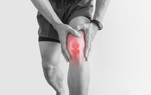
Joint pain (arthralgia) is a very common problem and may be related to infection or toxicity, trauma, inflammation, or cartilage degradation.
In most cases, joint pain will go away on its own within a few days. However, some situations require you to see a doctor as soon as possible. Even an experienced expert is not easy to determine the exact cause of joint pain, because early symptoms can be deceptive, and sometimes only 1-2 months or more can be fully understood.
The information in this article will help you understand the various diseases and conditions that cause joint pain. Modern diagnostic methods will enable you to determine the exact cause of the disease and choose the correct treatment strategy with your doctor.
In this article, we will study the injuries of multiple joints throughout the body. Sometimes a person starts to get hurt, and other joints will soon join his ranks. Over the course of a few days or weeks, the pain seems to shift from one part of the body to another. Many diseases cause pain in a group of joints in the form of seizures-epileptic seizures, when the pain subsides and then reappears.
Joint pain caused by a viral infection
In most cases, joint pain occurs with various viral infections: due to the direct effect of the virus on the joints or under the influence of toxins that accumulate in the blood during the acute phase of many infectious diseases.
In most cases, pain occurs in the small joints of the arms and legs, knee joints, and sometimes in the spine joints. The pain is not strong, very painful. It is called joint pain. Mobility is usually unaffected, and there is no swelling or redness. In some cases, a rash similar to urticaria may appear, but it will disappear soon. In most cases, viral joint pain becomes the first symptom of discomfort and is accompanied by fever, muscle pain, and weakness.
Despite the deterioration of general health, joint pain caused by viral diseases usually does not cause serious concern. Symptoms can be relieved by taking non-steroidal anti-inflammatory drugs, drinking plenty of water and rest. A few days later, the pain disappeared and the joint function was fully restored. There is no irreversible change in the structure of the joint.
Viral joint pain is a characteristic of, for example, influenza, hepatitis, rubella, mumps (adults).
Reactive arthritis
This is a group of diseases in which joint pain occurs after infection with viruses and bacteria. The direct cause of reactive arthritis is a fault in the immune system, which can cause inflammation of the joints, even though they are not affected by the infection.
Acute respiratory infections, intestinal infections, or genitourinary system diseases (such as urethritis or genital infections) are more common in joint pain 1-3 weeks. Unlike viral joint pain, joint pain is severe, accompanied by edema and impaired mobility. Body temperature may rise. Arthritis usually begins with the involvement of a knee or ankle joint. Within 1-2 weeks, the joints of the other half of the body begin to ache, and the small joints of the arms and legs begin to ache. Sometimes the joints of the spine are painful.
Joint pain usually disappears with treatment or on its own, leaving no sequelae. However, some types of reactive arthritis are chronic and occasionally worsen.
Wright's disease-A reactive arthritis that occurs after the transfer of chlamydia, which can take a chronic course. Joint pain in Reiter's disease usually precedes urinary dysfunction-a manifestation of chlamydial urethritis (inflammation of the urethra) and is usually overlooked. Then there are eye problems and conjunctivitis develops. If treatment is required, you must consult a doctor.
Reactive arthritis can develop after adenovirus infections, genital infections (especially chlamydia or gonorrhea), intestinal infections related to Salmonella, Klebsiella, Shigella, etc.
Joint pain when cartilage wears out
Diseases that accompany the gradual wear and tear of cartilage on the surface of the bones and joints are called degeneration. They are more common in 40-60 years and older, but they also occur in young people, such as those with joint injuries, professional athletes who exercise regularly, and obese people.
Deformed osteoarthritis (osteoarthritis, DOA)-This is a disease of the large joints of the legs-the knees and hip joints bear most of the load when walking. The pain comes gradually. After taking a break in the morning, the physical condition improves, and after a long time of walking and running in the evening and evening, the condition worsens. Inflammatory changes: Edema and redness are usually not obvious, and only appear in advanced cases. But people often complain of crackling in the joints. Over the years, the disease has continued to develop. Proteus joint disease is almost impossible to cure, it can only slow down the destruction of cartilage. In order to restore mobility, they resorted to surgery.
Spine condyleIt is another common degenerative disease. The reason is the thinning and destruction of cartilage between the vertebrae. The reduction in cartilage thickness causes compression of the nerves extending from the spinal cord and blood vessels. In addition to spinal joint pain, it can also cause many different symptoms. For example: headache, dizziness, arm and shoulder joint pain and numbness, heart, chest pain and interruption, leg pain, etc. Neurologists usually handle the diagnosis and treatment of osteochondrosis.
Autoimmune disease causes joint pain
Autoimmune diseases are a large group of diseases, the cause of which is not fully understood. All these diseases are combined by the characteristics of the immune system: the cells of the immune system begin to attack their own body tissues and organs, causing inflammation. Compared with degenerative diseases, autoimmune diseases are more likely to occur in childhood or young people. Their first manifestation is usually joint pain.
Joint pain is usually unstable: one joint pain today, another tomorrow, the day after tomorrow-the third. Joint pain is accompanied by edema, redness of the skin, joint movement disorders, and sometimes fever. After a few days or weeks, the joint pain disappears, but it will recur after a while. Over time, the joints will deform significantly and lose their mobility. A feature of autoimmune joint inflammation is morning stiffness. In the first morning, the affected joint must be kneaded for 30 minutes to 2-3 hours or more. The stronger the load on the joints the day before, the more time you need to spend warming up.
Gradually, symptoms of damage to other organs will add to joint pain: heart, kidneys, skin, blood vessels, etc. If left untreated, the disease will progress. It is impossible to cure it, but modern drugs can slow down this process. Therefore, the sooner the treatment is started, the better the effect.
Rheumatoid arthritis is the most common autoimmune disease and mainly affects the joints: they begin to suffer severe pain, redness and swelling. In most cases, the disease starts with pain in the small joints of the arms and legs: the joints of the fingers, hands, or feet, and less often-a failure of a knee, ankle, or elbow, followed by pain in other parts of the body.
Systemic lupus erythematosus-A rarer disease that is more likely to infect young women. It is characterized by flying pains in various joints of the body, deformed fingers, rashes on the skin, especially facial features-red forehead and cheeks, like butterfly wings. Joint pain may be accompanied by interruption and discomfort of the heart and chest, low-grade fever, weakness, weight loss, increased blood pressure, back pain, and edema.
Ankylosing spondylitis-Unlike lupus, it affects men more often. The disease begins with pain in the joints of the spine, waist, sacrum, and pelvis. Gradually, the pain spread upward to other parts of the spine. In addition to pain, stiffness, decreased flexibility, and over time, gait disturbances and complete immobility of the spinal joints are also characteristic. In the initial stage, ankylosing spondylitis can easily be confused with osteochondrosis. However, the first disease occurs in young men, and the second disease occurs in elderly people. As a diagnostic test, an X-ray is performed on the sacroiliac joint (the junction of the spine and pelvis). Based on the results of the study, doctors can confirm or deny the diagnosis.
Psoriasis joint pain
Psoriasis is a skin disease in which a characteristic rash appears on the surface of the body. Sometimes psoriasis affects the joints. The joints of hands and feet, fingers and feet, rarely the spine, are usually injured and swollen. A distinctive feature of psoriatic arthritis is asymmetric lesions. The skin on the joints may be blue-purple and can damage the nails. Over time, joint deformities and subluxations (the fingers begin to bend in an atypical direction).
Rheumatic joint pain
Rheumatism (acute rheumatic fever) is a serious disease caused by streptococcus. Rheumatism is characterized by very severe pain in the large joints of the legs and arms, which can appear 2-3 weeks after a sore throat or scarlet fever. It is more common in children. The pain is so severe that you cannot touch the joints and cannot move. The joints are swollen, red, and the temperature rises. First, some joints are injured, then other joints, usually symmetrical. Even without treatment, the pain will disappear on its own and the joint function will be fully restored. However, after a period of time, symptoms of severe heart damage will appear. Rheumatism requires urgent medical attention. Only timely treatment can avoid damage to the heart and other organs.
How to check for joint soreness?
There are many ways to check for joint pain. Usually, they are used in combination.
blood test-It is one of the most common tests for joint pain complaints. With the help of this research, it is possible to determine the presence of inflammation or indicate the degeneration of the disease, identify signs of infection, and use immunological tests or polymerase chain reaction (PCR) methods to accurately determine the presence of infectious or reactive arthritisThe pathogen of the disease in the case. The blood test showed a possible metabolic disorder, the state of internal organs.
Synovial fluid research-Clean the liquid on the joint surface. With its help, the articular surface is nourished and the friction during exercise is also reduced. Based on the composition of synovial fluid, the laboratory assistant draws conclusions about whether there is inflammation or infection in the joints, cartilage destruction and nutritional processes, and salt accumulation that can cause pain (such as gout). A needle is used to extract synovial fluid for analysis, and the needle is inserted into the joint cavity after local anesthesia.
Combined X-ray and computed tomography (CT)-A method that allows you to consider the structure of the bones of the joints, and indirectly judge the condition of the cartilage by the size of the joint space-the distance between the bones in the joint. X-ray examination is one of the preferred methods for joint pain. X-rays show mechanical damage to bones (fractures and cracks), joint deformities (subluxation and dislocation), bone growth or defect formation, bone density, and other criteria that help doctors determine the cause of joint pain. Computed tomography is also a method of X-ray research. Through a CT scan, the doctor will receive a series of layer-by-layer pictures of the joints, which in some cases can provide more complete information about the disease.
Joint ultrasound and MRI-The method is different in nature, but the purpose is similar. With the help of ultrasound or magnetic resonance imaging, information about the condition of joints and cartilage soft tissue can be obtained. Ultrasound and MRI show the thickness of the cartilage, its defects, and the presence of foreign inclusions in the joints. It also helps determine the viscosity and amount of synovial fluid.
Arthroscopy-A method of visual inspection of joints using microsurgery instruments, which are introduced into the cavity of the diseased joint after anesthesia. During the arthroscopy, the doctor has the opportunity to examine the internal structure of the joint with eyes, pay attention to its damage and changes, and take fragments of the joint synovium and other structures for analysis. If necessary, after the examination, the doctor can immediately perform the necessary treatment operations. Everything that happens during the arthroscopy is recorded on a disk or other storage medium, so you can consult other experts after the operation.
Combination therapy
If you have joint pain, find a good therapist or pediatrician for the child. He will make an initial diagnosis and refer you to a specialist for treatment if necessary. If joint pain is related to arthropathy or arthritis, treatment is likely to be handled by the rheumatologist here.
If the cause of joint pain is inflammation, use drugs to treat joints that can reduce inflammation. First of all, these are non-steroidal anti-inflammatory drugs (NSAIDs): indomethacin, ibuprofen, diclofenac, nimesulide, meloxicam and many others. If these drugs are not effective enough, the drugs in the corticosteroid group are prescribed in the form of injections into the joint cavity or tablets. When infection causes pain, antibiotics are given.
Special treatment programs are used for autoimmune diseases. For continuous hospital admissions, doctors choose the smallest effective dose of drugs that can strongly inhibit the inflammatory response or suppress the immune system. For example: sulfasalazine, methotrexate, cyclophosphamide, azathioprine, cyclosporine, infliximab, rituximab, etc.
For joint degenerative diseases (osteochondrosis, osteoarthritis), there are no specific drugs. Treatment of diseased joints includes prescribing anti-inflammatory and analgesics during the deterioration period, and taking metabolites based on chondroitin sulfate and hyaluronic acid. Although the effectiveness of the latter has not been recognized by all doctors.
If joint function deteriorates irreversibly, they will turn to surgery. Currently, there are a variety of endoprosthetic methods that allow implantation of artificial joints or parts thereof, rather than damaged or worn joints.